Computed Tomography (CT) Scan Reads From $40 Per Study
High Quality And Accurate Computed Tomography (CT) Scan Reads And Interpretations
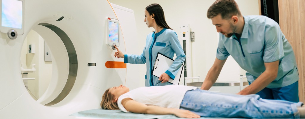
A computed tomography (CT) scan is a safe, quick imaging test to show layers of the body’s structures in great detail.
NDI provides CT (computed tomography) imaging scan interpretations, CT reading services, CT study reporting and CT secondary reads via teleradiology in all 50 states. Computed tomography, more commonly known as a CT or CAT scan, is a diagnostic medical imaging test.
To request rates for CT interpretations, please call 216-514-1199 or complete this email form.
Request A CT Scan Interpretation Or Computed Tomography Reading And Reporting Services
Computed Tomography (CT) Scan Reading Services, Interpretations And Reporting Services From NDI
About NDI CT Reading Services
NDI provides CT scan reading services via teleradiology in all 50 states. Remote CT reading and diagnostic image interpretation service prices start at $40 per study dependent upon volume. NDI radiology reading fees start at $12 per study.
NDI radiologists read CT scans that are used when imaging complex bone fractures, severely eroded joints, or bone tumors because they usually produces more detail than would be possible with a conventional x-ray.
NDI reads and interprets computed tomography scans and imaging for people and groups throughout the United States. Computed tomography (CT) imaging provides a form of imaging known as cross-sectional imaging.
The fellowship trained and US board certified NDI teleradiologists provide final radiology reads, outside reads and radiology second reads to our customers. Computed tomography, more commonly known as a CT or CAT scan, is a diagnostic medical imaging test.
A CT scan generates images that can be reformatted in multiple planes. It can even generate three-dimensional images. Doctors can review these images on a computer monitor, print them on film or via a 3D printer, or transfer them to a CD or DVD.
NDI provides CT interpretations and radiology image interpretations via teleradiology. The easiest way to get access to teleradiology services in the United States is to call National Diagnostic Imaging at 1-800-950-5257 or email info@ndximaging.com.
What Is A Computerized Tomography (CT) Scan?
Computed tomography, or CT, scans are medical imaging tests that use ionizing radiation to create cross-sectional (slices) pictures inside selected areas of the body from different angles.
The images can show internal organs, blood vessels, soft tissues, and bones. CT scans combine a series of x-ray (radiography) images into a three-dimensional picture. Some CT procedures may use a contrast dye which allows healthcare providers to see specific organ(s).
A CT scan uses X-rays and computers to produce images of a cross-section of your body. When interpreting at CT scan, it is important to determine the orientation.
It takes pictures that show very thin “slices” of your bones, muscles, organs and blood vessels so that healthcare providers can see your body in great detail.
A computerized tomography (CT) scan combines a series of X-ray images taken from different angles around your body and uses computer processing to create cross-sectional images (slices) of the bones, blood vessels and soft tissues inside your body. CT scan images provide more-detailed information than plain X-rays do.
Healthcare providers ask patients for a CT scan to detect, diagnose, or plan treatment for a particular disease or injury.
Common uses of CT scan include checking for:
- Tumors
- Infections
- Blood clots
- Internal bleeding
NDI Reads And Interprets CT Scans For Individual Patients And Imaging Centers
Because of COVID-19, many radiological practices have embraced teleradiology. When the COVID-19 pandemic hit in early 2020, radiology practices and departments started to increase their use of teleradiology and virtual technology tools to maintain their workloads from remote locations.

Chest CT has a potential role in the diagnosis, detection of complications, and prognostication of coronavirus disease 2019 (COVID-19). Chest CT is valuable to detect both alternative diagnoses and complications of COVID-19 (acute respiratory distress syndrome, pulmonary embolism, and heart failure), while its role for prognostication requires further investigation.
This study aimed to assess the characteristic chest X-ray features of COVID-19 and correlate them with clinical outcomes of patients. Chest CT imaging may be used to stratify the severity of lung involvement and to predict outcomes in COVID-19, which in turn may assist physicians with proper triaging of patients and allocation of resources.
Knowledge of the natural temporal evolution of lung abnormalities in COVID-19 may be helpful to radiologists in determining the stage of disease and in distinguishing them from potential complications when evaluating chest CT examinations.
Computed tomography imaging studies are read by National Diagnostic Imaging on behalf of imaging centers, private individuals, physicians, radiologists, hospitals, office-based imaging practices and outpatient clinics. Mobile imaging studies are welcomed, and we take the time to provide accurate detailed reports.
National Diagnostic Imaging prides itself in providing customized turn-around times for routine, priority, and STAT reports (critical/urgent) at no additional charge. Daytime and nighttime CT reading services are available.
Remote CT reading and diagnostic image interpretation service prices start at $33 per study dependent upon volume.
NDI Provides The Following Types Of CT Scan Interpretations, CT Reads And Computed Tomography Diagnostic Imaging Reporting Services In All 50 States
- CT Chest Scan Interpretations, Radiology Reads And Image Diagnostic Reporting Services
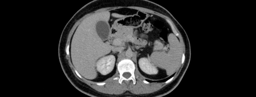
- CT Abdomen Reads And Interpretations
- CT Pelvis Reads And Interpretations
- CT Chest/Abdomen/Pelvis Interpretations (Full-Body) Reads And Interpretations
- CT Abdomen/Pelvis Reads And Interpretations
- CT Brain Reads And Interpretations
- CT MSK (Musculoskeletal) Reads And Interpretations
- CT Lumbar Reads And Interpretations
- CT Cervical Reads And Interpretations
- CT Thoracic Reads And Interpretations
- CT Head Reads And Interpretations
- CT Neck Reads And Interpretations
- CT Breast Reads And Interpretations
- CT Upper Extremity Reads And Interpretations
- CT Lower Extremity Reads And Interpretations
- CT Urogram Reads And Interpretations
If your organization is looking for additional information and or a teleradiology solution for your CT interpretations, please contact National Diagnostic Imaging.
How to Read a CT Scan Chest – A Radiologists Approach
Posted On YouTube On March 3, 2021 by Medical Basics
Computed tomography (CT) is a diagnostic imaging test used to create detailed images of internal organs, bones, soft tissue and blood vessels.
The cross-sectional images generated during a CT scan can be reformatted in multiple planes, and can even generate three-dimensional images which can be viewed on a computer monitor, printed on film or transferred to electronic media.
CT scanning is often the best method for detecting many different cancers since the images allow your doctor to confirm the presence of a tumor and determine its size and location.
CT is fast, painless, noninvasive and accurate. In emergency cases, it can reveal internal injuries and bleeding quickly enough to help save lives.
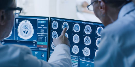
NDI Radiologists Read And Interpret Computed Tomography (CT or CAT) Scans of the Brain
A CT of the brain is a noninvasive diagnostic imaging procedure that uses special X-rays measurements to produce horizontal, or axial, images (often called slices) of the brain.
Brain CT scans can provide more detailed information about brain tissue and brain structures than standard X-rays of the head, thus providing more data related to injuries and/or diseases of the brain.
Computed tomography (CT) of the head uses special x-ray equipment to help assess head injuries, severe headaches, dizziness, and other symptoms of aneurysm, bleeding, stroke, and brain tumors. Learn how to read a head CT, here.
How to Read a CT Scan of the Head – MEDZCOOL
Posted On YouTube On May 1, 2016 by Medzcool
NDI Radiologists Read Computed Tomography (CT) Scans To Detect Tumors And Cancer
Doctors at National Diagnostic Imaging use a computed tomography (CT) scan, also called a CAT scan, to find cancer.
A CT scan takes pictures of the inside of the body using x-rays taken from many angles. A computer combines these pictures into a detailed, 3-dimensional image. This image will show abnormal areas and any tumors.
A computed tomography scan — also called a CT or CAT scan — is an imaging test that lets doctors see inside a person’s body.
As a patient lies still on a table, special X-ray equipment takes pictures from different angles to create cross-sections of the body.
The “slices” can be looked at individually or together to create 3-D views of tissues and organs, according to the National Cancer Institute. The test is painless and usually done on an outpatient basis.
A computed tomography (CT) scan can create cross-sectional images of any part of your body using special X-rays and a computer.
This type of radiology study is an important part of diagnosing medical diseases like strokes, cancers, and infections in the abdomen like appendicitis.
You can learn to read a CT scan if you understand the normal anatomy and what the shades of white, grey, and black on the films mean.
CT Head Interpretation
The CT head scan is a computer-generated series of images from multiple X-rays taken at different levels. Fine X-ray beams passed through the subject are absorbed to different degrees by different tissues and the transmitted radiation is measured by a scanning device.
- Interpretation: Use a systematic approach to viewing the anatomical structures in the many slices prepared by the CT scanner.
- Blood
- Cisterns
- Brain Matter
- Ventricles
- Bone
What Does My CT Scan Tell?
CT scans show a slice, or cross-section, of the body. The image shows your bones, organs, and soft tissues more clearly than standard x-rays.
CT scans can show a tumor’s shape, size, and location. They can even show the blood vessels that feed the tumor – all without having to cut into the patient.
A Systematic Approach To The Interpretation Of CT Head
A systematic approach to the interpretation of CT (computed tomography) of the head for the emergency physician.
The emergency physician should be adept at the interpretation of computed tomography of the head, particularly for life-threatening processes where awaiting a radiologist interpretation may unnecessarily delay care.
Contact National Diagnostic Imaging For CT Reading Services
Please contact National Diagnostic Imaging by calling (216)-514-1199, by emailing info@ndximaging.com or complete the form below to get a quote for computed tomography interpretation services.
Accreditation Programs For Diagnostic Imaging Centers In The U.S.

Diagnostic imaging lets doctors look inside human and animal bodies for clues about a medical condition. A variety of machines and techniques can create pictures of the structures and activities inside the body. The type of imaging a doctor uses depends on the symptoms and the part of your body being examined. Ultrasonography is a popular diagnostic imaging tool that looks inside a dog or cat’s body via the use of sound waves.
ACR Accreditation is recognized as the gold standard in medical imaging.
The ACR offers accreditation programs in CT, MRI, breast MRI, nuclear medicine and PET as mandated under the Medicare Improvements for Patients and Providers Act (MIPPA) as well as for modalities mandated under the Mammography Quality Standards Act (MQSA).
Accreditation application and evaluation are typically completed within 90 days.
The ACR has accredited more than 39,000 facilities in 10 imaging modalities. They offer accreditation programs in Mammography, CT, MRI, Breast MRI, Nuclear Medicine and PET, Ultrasound, Breast Ultrasound and Stereotactic Breast Biopsy.
The Joint Review Committee on Education in Radiologic Technology (JRCERT) accredits educational programs in radiography, radiation therapy, magnetic resonance, and medical dosimetry.
The National Accreditation Program for Breast Centers (NAPBC) provides the structure and resources you need to develop and operate a high-quality breast center.
Programs that are accredited by the NAPBC follow a model for organizing and managing a breast center to facilitate multidisciplinary, integrated, comprehensive breast cancer services.
Get information from the Centers for Medicare & Medicaid Services (CMS) about their requirements for accreditation of advanced diagnostic imaging suppliers, here.
The Intersocietal Accreditation Commission (IAC) is a nonprofit, nationally recognized accrediting organization.
The IAC was founded by medical professionals to advance appropriate utilization, standardization and quality of diagnostic imaging and intervention-based procedures.
The IAC is a nonprofit organization in operation to evaluate and accredit facilities that provide diagnostic imaging and procedure-based modalities, thus improving the quality of patient care provided in private offices, clinics and hospitals where such services are performed.
With a 30-year history of offering medical accreditation to facilities within the U.S. and Canada, IAC is also now offering accreditation in international markets.
The IAC programs for accreditation are dedicated to ensuring quality patient care and promoting health care and all support one common mission: Improving health care through accreditation®.
The ACVR is the American Veterinary Medical Association (AVMA) recognized veterinary specialty organization™ for certification of Radiology, Radiation Oncology and Equine Diagnostic Imaging.
If you are a radiology imaging service in the United States that is looking for a company that can provide teleradiology coverage for your current and future case volume, contact National Diagnostic Imaging by phone at 216-514-1199 or by emailing info@ndximaging.com.